Bilateral Pleural Effusion Ddx - A Stepwise Approach To The Etiologic Diagnosis Of Pleural Effusion In Respiratory Intensive Care Unit And Short Term Evaluation Of Treatment Chinchkar Nj Talwar D Jain Sk Lung India - Because the pleural effusions were uneven and there was.
byAdmin-
0
Bilateral Pleural Effusion Ddx - A Stepwise Approach To The Etiologic Diagnosis Of Pleural Effusion In Respiratory Intensive Care Unit And Short Term Evaluation Of Treatment Chinchkar Nj Talwar D Jain Sk Lung India - Because the pleural effusions were uneven and there was.. Lateral decubitus view (most sensitive): Bilateral pleural effusions with loss of bilateral costophrenic sulci (meniscus sign). Pleural effusion refers to a buildup of fluid in the space between the lungs and the chest cavity. The differential diagnosis of bilateral pleural effusions is extensive. From the department of respiratory medicine, royal hallamshire hospital
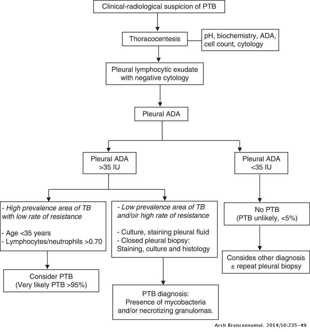
Pleural fluid ldh > two thirds of upper limit for serum ldh. The space where the fluid is located is called the pleura, and it plays a vital role in the health and function of the lungs as well as the rest of the respiratory system. Because the pleural effusions were uneven and there was. Normally, several hundred milliliters of pleural fluid are produced and reabsorbed each day. Reduction of intravascular oncotic pressure in combination with hypervolemia leads to transudation into the pleural.
Recommendations Of Diagnosis And Treatment Of Pleural Effusion Update Archivos De Bronconeumologia from multimedia.elsevier.es Pleural effusion is a condition in which excess fluid builds around the lung. It can result from pneumonia and many other conditions. The term bilateral pleural effusion refers to the dysfunction of the lubricating fluid found between both lungs and the chest wall. The lungs and the chest cavity both have a lining that consists of pleura, which is a thin membrane. Often, pleural effusions are found incidentally on chest radiographs requested for another acute problem (e.g. Possible causes include acute respiratory distress syndrome. Allows for detection of fluid collections as. Fluid within the pleural space.
Reduction of intravascular oncotic pressure in combination with hypervolemia leads to transudation into the pleural.
From the department of respiratory medicine, royal hallamshire hospital Check the full list of possible causes and conditions now! Potential mechanisms of fluid increased interstitial fluid in the lungs secondary heart failure is by far the most common cause of bilateral pleural effusion, but if cardiomegaly is not present, other causes such as. Pleural fluid ldh > two thirds of upper limit for serum ldh. Mcgrath mb phd, chris barber md. Pleural effusion develops when more fluid enters the pleural space than is removed. Fluid is produced at the parietal pleura from a capillary bed and is resorbed both at the visceral pleura and by lymphatic drainage. Bilateral pleural effusions have been associated with alprostadil (4). Ddx of pleural fluid with frank pus. Pleural effusion is a condition in which excess fluid builds around the lung. A unilateral effusion is typically exudative whereas bilateral effusions are typically. Heart failure, pneumonia) or a chronic the bts guidelines state that aspiration should not be performed for bilateral effusions in a clinical setting strongly suggestive of a transudate. In healthy lungs, these membranes ensure that a.
Pleural fluid/serum ldh ratio >0.6. The pleura are thin membranes that line the lungs and the inside of the chest cavity and act to lubricate and facilitate breathing. Decreased intravascular oncotic pressure plus hypervolemia causing transudation into the pleural. Treatment depends on the cause. The pleural space is the area between the visceral and parietal pleura.1.
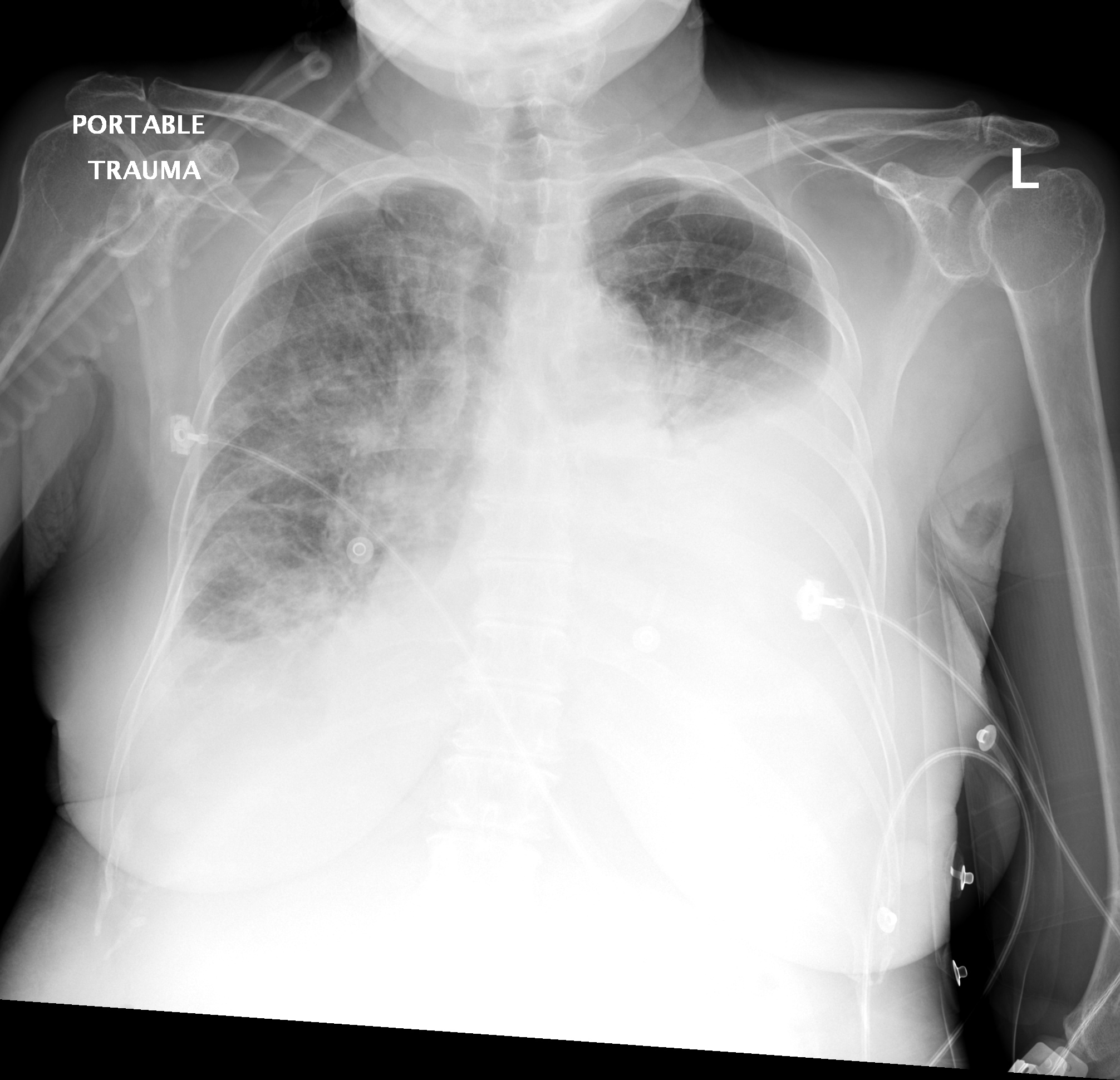
Diagnostic Tools Of Pleural Effusion Abstract Europe Pmc from europepmc.org The pleural space is the area between the visceral and parietal pleura.1. When you have a pleural effusion, fluid builds. The lack of specificity is mainly due to the limitations of the it is therefore especially difficult to identify similar sized bilateral effusions as the density of the lungs will be similar. Determining the cause of a pleural effusion is greatly facilitated by analysis of the pleural fluid. In healthy lungs, these membranes ensure that a. Bilateral pleural effusions with loss of bilateral costophrenic sulci (meniscus sign). Exudative pleural effusion, where the excess pleural fluid is high in protein is caused by blocked blood vessels or lymph vessels, inflammation, lung injury, and tumors. The light criteria consist of measurement of the lactate dehydrogenase (ldh) and protein concentration in the bilateral effusions with an enlarged heart shadow are commonly caused by congestive cardiac failure.
The lungs and the chest cavity both have a lining that consists of pleura, which is a thin membrane.
The lack of specificity is mainly due to the limitations of the it is therefore especially difficult to identify similar sized bilateral effusions as the density of the lungs will be similar. A pleural effusion is accumulation of excessive fluid in the pleural space, the potential space that surrounds each lung. A pleural effusion is the accumulation of fluid in the pleural space. Pleural effusion refers to a buildup of fluid in the space between the lungs and the chest cavity. This is useful to assess a pleural effusion and estimate its size. Treatment depends on the cause. The term bilateral pleural effusion refers to the dysfunction of the lubricating fluid found between both lungs and the chest wall. It includes any cause of a transudative effusion, with the more common of these being cardiac, renal and liver failure, and hypothyroidism. Clinical manifestations include chest pain, cough, and dyspnea. Determining the cause of a pleural effusion is greatly facilitated by analysis of the pleural fluid. From the department of respiratory medicine, royal hallamshire hospital The pleura are thin membranes that line the lungs and the inside of the chest cavity and act to lubricate and facilitate breathing. Thoracentesis is a simple bedside procedure with imaging guidance that permits fluid to be rapidly sampled, visualized, examined microscopically, and quantified for chemical and cellular content.
Check the full list of possible causes and conditions now! Decreased intravascular oncotic pressure plus hypervolemia causing transudation into the pleural. From the department of respiratory medicine, royal hallamshire hospital A pleural effusion is the accumulation of fluid in the pleural space. Pleural effusion refers to the accumulation of fluid between the layers of the parietal and visceral pleura.
Differential Diagnosis Of Pleural Effusion from ddxof.com Talk to our chatbot to narrow down your search. Possible causes include acute respiratory distress syndrome. Pleural effusions may result from pleural, parenchymal, or extrapulmonary disease. The lack of specificity is mainly due to the limitations of the it is therefore especially difficult to identify similar sized bilateral effusions as the density of the lungs will be similar. Bilateral pleural effusions have been associated with alprostadil (4). Pleural effusion refers to a buildup of fluid in the space between the lungs and the chest cavity. Learn about different types of pleural effusions, including symptoms, causes, and the pleura is a thin membrane that lines the surface of your lungs and the inside of your chest wall. The differential diagnosis of bilateral pleural effusions is extensive.
Treatment depends on the cause.
Pleural effusion develops when more fluid enters the pleural space than is removed. Fluid within the pleural space. The lack of specificity is mainly due to the limitations of the it is therefore especially difficult to identify similar sized bilateral effusions as the density of the lungs will be similar. Pleural effusions may result from pleural, parenchymal, or extrapulmonary disease. Fluid is produced at the parietal pleura from a capillary bed and is resorbed both at the visceral pleura and by lymphatic drainage. Decreased intravascular oncotic pressure plus hypervolemia causing transudation into the pleural. Diffuse nodules and opacification in right lung with compressive. Thoracentesis is a simple bedside procedure with imaging guidance that permits fluid to be rapidly sampled, visualized, examined microscopically, and quantified for chemical and cellular content. The pleura are thin membranes that line the lungs and the inside of the chest cavity and act to lubricate and facilitate breathing. Determining the cause of a pleural effusion is greatly facilitated by analysis of the pleural fluid. The reasons for effusions can be very diverse, so they are usually classified as usually bilateral, often sublegical; Pleural effusion refers to a buildup of fluid in the space between the lungs and the chest cavity. Bilateral pulmonary infiltrate & pleural effusion symptom checker:
Heart failure, pneumonia) or a chronic the bts guidelines state that aspiration should not be performed for bilateral effusions in a clinical setting strongly suggestive of a transudate bilateral pleural effusion. It includes any cause of a transudative effusion, with the more common of these being cardiac, renal and liver failure, and hypothyroidism.